Segment structure
As well as plant life, Amthalassa is inhabited by hydrogen
breathing animal life. The physically largest and most prominent group of
animals are those belonging to the phylum Plurafistulata.
They are characterised by the basic arrangement of their
bodies, with multiple tube-shaped segments lined up together. Early in their
evolution these tubes were almost identical to each other, but over time they
became more specialised. Plurafistulates have radial symmetry, and each layer of
tube segments starting from the central segment serves a specific role.
The basic form that each segment used to have is still found
in many simple, single segmented animals alive today, descended from the same
ancestor. In the centre of such tubes is a digestive tract, with nerves and two
major blood vessels further out running across the length of the tube. These
blood vesicles are surrounded in muscle that allows it to contract in a motion
similar to that of a digestive tract to draw blood across, and as it does so
some of this blood is forced into smaller capillaries. Around all of this is a
layer of muscle tissue, and along the outside of the segment is epithelial
tissue.
As segments have become specialised in more complex life,
however, most of these features have become reduced, although the needed
features have increased in complexity. The digestive tract, for example, is
only present in the central segment of most organisms within the phylum Plurafistulata, and muscular sections of the veins and arteries of the circulatory
segments have developed to the point that they have become hearts. In simpler
organisms consisting of a single tubular segment, there is very little
distinction between the cross section of one part of a tube and another.
Development
One of the simplest forms of plurafistulate are boneless fish-like
animals that can be found in the ammonia oceans of the planet. Most are
limbless and serpentine in appearance, and possess a ring of eyes radially
surrounding the mouth at the front of their head. Many species have developed
an extension of the muscle segments at the front of their mouths that have
eventually formed tentacles for use in the manipulation of food. Some have
moved away from radial symmetry by developing fins along the tops and bottoms
of their bodies, with some even possessing tail fins. However, it is the more
snake-like, radially symmetrical fish that segmented land animals evolved from.
The first plurafistulates on land, having evolved from limbless
fish, moved in a snakelike fashion, and many similar amphibious animals still
exist today. Without oxygen, Amthalassa lacks an ozone layer, and as such the
surface is exposed to high amounts of UV radiation in modern times. However, since
the hazy tholin layer in the upper atmosphere was very thick at this point in
the planet’s past – a product of the interaction of atmospheric methane with
sunlight – these creatures needed very little protection against solar
radiation. This layer blocked out not only UV light but also a great deal of
the visible spectrum, making the planet a fairly dim place at this point in
history. Weak against sunlight, these animals tended to retreat to darker areas
at points in history when this tholin layer was thinner, and even today their
most basal descendants still prefer darkness.
Since Amthalassa has usually been quite forested, and was
especially forested during the damp period of history when plurafistulates first
moved to land, many animals took to the trees (plants during this period mainly
used infrared radiation for photosynthesis (although they moved toward the visible
spectrum later on), so the tholin layer wasn’t as much of an issue for them as
it would otherwise be). While many tree “snakes” evolved during this period, some
species of the early snake-like amphibians adapted to use their facial
tentacles for locomotion. As their tentacles elongated, these tree dwelling
amphibians became more octopus-like in appearance.
Most large land animals evolved from a group of these
tree-octopuses that developed a hard exoskeleton as a response to a much drier
and brighter point in the planet’s history. This clade is called Exoskeletida. With
this exoskeleton, they were able to walk on land using what were once their
tentacles as reasonably sturdy legs.
These exoskeletons allowed this group to quickly diversify
since, in addition to the support it gave their bodies, their exoskeletons also
allowed them to survive in a wide range of conditions, regardless of the amount
of light they were exposed to or the level of humidity. They were able to
survive in arid steppes just as they could survive in the now shrinking jungles
their ancestors were previously limited to; the lack of tree cover no longer
posed an issue and their exoskeletons protected them from losing moisture.
Exoskeletida anatomy
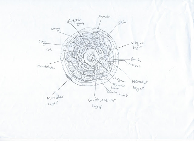
 |
| Vertical cross section of a typical exoskeletid |
Digestive system
The single innermost tube of an exoskeletid serves the
function of digestion. At the bottom of the tube is an oral opening. Many
species have developed inner tongues, a series of tentacle-like extensions of
the muscles of the central tube, for assistance in the swallowing of food. There
is a short oesophagus, with sphincters at either end, leading to a stomach
where food is broken down. Since ammonia is more alkaline than water, the
stomach acids don’t have to have as low a pH and even a pH of 7 is considered
acidic to life on Amthalassa. Above this is an intestine that stretches across
the tail present at the top of the animal’s body. The intestine eventually
leads to an anus or cloaca at the end of the tail.
The digestive organs are surrounded by a layer of energy
storing tissue which primarily contains alkynes, nitriles, and various
unsaturated hydrocarbons. There are two primary blood vessels running through
this, one vein and one artery, often with a number of tiny “heartlets” to
assist in the pumping of blood. There is one primary nerve in this segment,
which branches out into numerous smaller nerves. Outside of the alkyne tissue
is a thin layer of muscle, which is surrounded by epithelial tissue.
Cardiorespiratory system
Surrounding this innermost segment are a series of
cardiorespiratory segments, varying in number depending on species. At the
bottom of each segment is a lung, capable of drawing in and expelling air from
the contraction and relaxing of muscles in the muscular layer of the segment.
There are two entrances to the lungs; openings leading to the mouth and a
“nostril”. Each opening is connected to a different side of the lung, the
nostrils to the top of the lung and the one in the mouth to the bottom. The
nostril actually originally evolved from the gills at the sides of their
fish-like ancestors bodies, with ammonia entering through mouths of these fish
as they swim and flowing out through what is now their nostrils. Certain
species developed a pumping method to draw air into their mouths and out of
their gills in order to respire more effectively, some even going as far as using
these gill pumps as a means of locomotion via jet propulsion. It is these gill
pumps that later became the lungs of land animals, who can now breathe either
in or out from both openings.
Above the lungs are a series of hearts running along the
tube segment. The one closest to the lung is the primary heart, responsible for
pumping dehydrogenated blood towards the lungs and hydrogenated blood to the legs
and brain. The other hearts pump blood to the various organs and to muscle
outside the legs, such as the tail, and also pump venous blood back to the
primary heart.
This segment also contains a large, primary artery, as well
as a larger primary vein, both of which run through each heart. These are the
largest blood vessels of the body. Most larger organisms cannot obtain enough
energy purely from hydrogen dissolved in the blood plasma, and as such make use
of hydrogen carrying substances. In the case of most plurafistulates, this is an
iridium based compound similar to a substance called Vaska’s complex known to
Earth scientists. It is yellow or orange in colour and carried by blood cells.
Like all segments, there is a layer of muscle and then epithelial
tissue on the outside.
After hydrogen is breathed in, a mixture of methane, ethane,
and ammonia is exhaled as by-products of respiration.
Nervous system
Surrounding the cardiorespiratory segments are segments
containing the central nervous system. While all segments have one primary
nerve running through them, these segments have a much more developed one, the plurafistulate equivalent of a spinal cord. Early in the evolutionary history of Plurafistulata, the nerves at the bottom of these segments bunched together into
ganglia and then eventually evolved into a brain. In spite of the fact there
are multiple nervous segments, in the exoskeletid body there is only a single
brain; the brain tissue of each segment is connected to the brain tissue of
every other segment, so that it forms a single, ring shaped multi-lobed brain.
The epithelial tissue on the outside of the part of the
segment surrounding the brain is very hard and chitinous – and in some cases
has undergone ossification – in order to protect the brain. While not as hard
as the outer exoskeleton, this still serves to defend it against damage.
 |
| Horizontal cross section of an exoskeletid |
Muscular system
The outermost segments of exoskeletids are the muscular
segments. The muscle that originally surrounded each segment has become so
developed so as to fill the entire tube. This allows for movement of the tail,
and is especially important in non-exoskeletid plurafistulates that need it for
structural support. The snake-like amphibians depend on this tail muscle for
locomotion.
Like all segments, these muscle segments contain a major
vein and artery and a number of small heartlets. In the case of the muscle segment
heartlets, their pumping can be assisted by the movement of muscle during times
of intense activity.
These segments extend further than all other segments in exoskeletids and many other plurafistulates, beyond the
mouth, forming legs (or tentacles in the case of non-exoskeletids). In exoskeletids these legs are surrounded by jointed exoskeleton, providing support and
allowing the organism to walk. Since these legs are an extension of the muscle
segment, not only are they muscle filled, but they are also equal in number to
the amount of such segments the organism possesses. Many species possess toes
at the ends of these legs, which initially evolved when the tentacles of their
ancestors split and branched at the end for better grasping of trees or finer
manipulation of food.
On the outside is a second layer of alkyne, alkene and
nitrile tissue, surrounded by skin and then the exoskeleton. The exoskeleton is
composed primarily of a substance close to chitin as well as, in hardened
areas, calcium carbonate – although this calcium carbonate is part of a tissue
with a specific structure that allows it to become much harder.
Sensory organs
 |
| Structure of a typical plurafistulate eye |
The surface of their exoskeletons (as well as the skin of
non-exoskeletids) possesses numerous sensory hairs, allowing them to feel touch
even on the most armoured parts of their bodies. In many amphibious and fish
species some of the sensory hairs on their tentacles, where these hairs are
most concentrated (as the tentacles are used as feelers), have become sensitive
to vibrations. In exoskeletids the area around these hairs have become
increasingly concave for protection to the point that they are located within
an internal cavity. These holes, which are located on their feet, serve as
ears.
A line of eyes surrounds the bodies of exoskeletids, giving
them 360 degree peripheral vision in a horizontal plane. Of course, objects can
still be out of their sight above or below them, and the amount of vertical peripheral vision varies from
species to species. For example, those living in flat, mossy plains have very
little need for vertical peripheral vision, perfectly comfortable with their
ability to see clearly in every horizontal direction. However, vertical
peripheral vision tends to be high in species inhabiting dense jungles, where
tree dwelling organisms may be lurking above.
The number of eyes an animal possesses is usually equal to
the number of limbs, with one eye per outer segment.
The actual structure of the eye shares a degree of
similarity to the camera eyes of Earth and many other planets due to convergent
evolution. However, due to their independent development, there are a number of
significant differences. The lens, for example, is made of calcite, much like
the eyes of trilobites. There is not just one of these lenses, but two; one
outer and one inner. While human eyes focus light by changing the shape of the
lens, this is not possible to do with the harder calcite lenses of
plurafistulates. Instead, the eye focuses by changing the position of the lens, much like cephalopod eyes. However, it
functions differently to a cephalopod eye in that it is a second lens inside
the eye that moves. As this inner lens changes position relative to the outer
lens, the image changes focus. The inner lens is attached to a membrane
separating two halves of the eye, and its position is changed by a series of
muscles attached to it, in addition to openings in the membrane that allow
fluid inside the eye to move from one half to the other. Not only can the inner
lens move towards and away from the outer lens, but it can move slightly side
to side and change angle.
In most species, the actual eye itself isn’t attached to
muscle and cannot change position, but with 360 degrees peripheral vision many
species don’t need to move their eyes that much. Those that need to see above
and below themselves, for example species living in jungles, have to move their
bodies slightly to look around. Slight movement may also be done by mossland
species just to bring already visible objects into better focus, especially if
they’re nearby.
Mouths
Most species of exoskeletids have a sheet of skin and muscle
underneath their mouths to prevent food from falling out while it is being
chewed. This extends from their muscular segments. There is a hole in the
middle of this sheet of skin held shut by a muscular sphincter.
Inside the mouth are many tentacle-like tongues. The inner
tongues, mentioned earlier, are part of the digestive segment, and are used
primarily to assist in the swallowing of food. The outer tongues are radulae attached to the muscular segment and perform a wider range of functions; namely
the manipulation of food and for chewing. As the oral sphincter opens they
grasp food and bring it into the mouth, then begin grinding the food up with
the mouth closed using the many chitinous teeth on each tongue. The radulae also usually possess sensory hairs capable of distinguishing taste. Food is
often tasted before bringing it into the mouth in order to assess it for
nutritional value and to ensure what’s being eaten is edible.
Reproductive system
Most groups of exoskeletid are hermaphroditic and reproduce
sexually. Their sexual organs are located in their tail, within the central
digestive segment, although the uterus is lower down where the body is thicker
and there’s more room for it. There is a canal connecting the uterus to the
mouth which eggs pass through.
Many species mate by rubbing their cloaca together, although
there are some species that have developed penises in their tails. Reproduction
can only usually take place after mating; although hermaphroditic, very few
exoskeletid species can self-fertilise.
I like this blog. :D Why has no one else commented on it before?
ReplyDeleteI don't know, I don't think a lot of people have seen it.
ReplyDelete